HORMONES 2008, 7(3):243-250
DOI: 10.14310/horm.2002.1204
Address for correspondence:
Galina Kolesnikova, National Center for Endocrinology RAMS, Dmitriya Uljanova Str. 11, Moscow, Russia, 117036, Tel./Fax: 7(495)1264458, e-mail: kolesnikova@endocrincentr.ru
Received 14-01-08, Revised 10-05-08, Accepted 30-05-08
Abstract
OBJECTIVE: The purpose of this study was to evaluate the direct action of desmopressin (agonist of vasopressin) on the hypophysis and the three zones of the adrenal cortex in patients with different forms of hypercortisolism. DESIGN: Forty-three patients with hypercortisolism—21 with Cushing’s disease (14 females, 7 males), 11 with extrapituitary, ectopic tumours (5 females, 6 males), and 11 with ACTH-independent Cushing’s syndrome (6 females, 5 males)—were evaluated. The response of the pituitary and adrenal glands was assessed by measuring plasma levels of Adrenocorticotropic Hormone (ACTH), cortisol, aldosterone, and dehydroepiandrosterone sulfate (DHEAS) at baseline and at 15, 30, 60, 90, and 120 min after the administration of desmopressin. RESULTS: We observed two main modes of secretory response: (1) elevation of the ACTH level followed by a rise of one, two or all three adrenal steroids, and (2) ACTH-independent elevation of adrenal steroids in various combinations. CONCLUSION: In a number of patients with hypercortisolism, the adrenal cortex responded to desmopressin administration by enhanced synthesis and secretion of glucocorticoids (cortisol), mineralocorticoids (aldosterone), and adrenal androgens (DHEAS) without a concomitant rise in ACTH. These findings suggest the presence of “ectopic” vasopressin receptors in the human adrenal cortex.
Key words: Adrenal adenoma, Adrenocorticotropic Hormone, Aldosterone, Cortisol, Cushing’s syndrome, Dehydroepiandrosterone-sulfate, Desmopressin, Ectopic receptors, Ectopic V1/V2 receptors
INTRODUCTION
Clinical symptoms of Cushing’s syndrome are known to be closely associated with the enhanced secretion of hormones synthesized by the adrenal cortex, mainly cortisol. The most common cause of ACTH-dependent hypercortisolism is Cushing’s disease [(CD) about 80%], ACTH producing extrapituitary tumour (ECT) being a less frequent form (15%). ACTH-independent Cushing’s syndrome is rare and is due to a tumour or micronodular hyperplasia of the adrenal cortex (ACTH-ind).1 The incidence rate of Cushing’s syndrome caused by unilateral adrenal adenoma is estimated at 2 cases per 1,000,000 population per year. The incidence of CD is 3 times more frequent than primary adrenal lesion, i.e. 5-6 cases per 1,000,000 per year (9-10 cases together with ECT). There are large gender differences in the incidence of adrenal adenoma, which occurs 4 times more frequently in women than in men.1
Vasopressin (VP), like corticotropin-releasing hormone (CRH), is an important regulator of pituitary ACTH secretion. After binding of CRH and VP to their respective receptors in corticotrophs (R1 for CRH and V3 for VP), the synthesis of proopiomelanocortin (POMC), a precursor of ACTH, is stimulated.2 Moreover, VP has been reported to exert direct action on neoplasias of the adrenal cortex where it activates the so-called “ectopic receptors”.3
The present study was designed to elucidate the direct effect of the VP agonist desmopressin (DM) on each of the three functional zones of the human adrenal cortex. To this end, we evaluated pituitary response (by measuring ACTH level) and the response of the zona glomerulosa, zona fasciculate, and zona reticularis (by measuring aldosterone, cortisol, and dehydroepiandrosterone-sulfate concentrations, respectively) to the action of DM in patients with different forms of hypercortisolism (Endogenous Cushing’s Syndrome).
SUBJECTS AND METHODS
The study group included 43 patients with hypercortisolism and 11 healthy individuals who served as controls.
Three groups of patients with hypercortisolism, aged 22-49 years, were studied during the active phase of the disease: 21 patients had Cushing’s disease (CD, 14 females and 7 males), 11 had ectopic (pulmonary), ACTH-producing tumour (ECT, 5 females and 6 males), and 11 had cortisol secreting adrenal tumour (ACTH-ind, 6 females and 5 males). In all patients the diurnal rhythm of ACTH and cortisol surges were examined (blood samples were taken at 08:00, 12:00, 16:00, and 23:00). Based on the 4 individual values the mean daily value was calculated.
The presence of pituitary adenomas and adrenal tumours was confirmed by magnetic resonance imaging (MRI), and the ectopic pulmonary ACTH-producing tumour by computed tomography (CT). After the surgical removal, the type of the tumours was confirmed histologically and immunochemically with antibodies to ACTH, where appropriate.
The control group consisted of 11 healthy subjects, aged 22 to 49 years: 7 females in the follicular phase of the menstrual cycle, and 4 males. In the controls, the diurnal rhythm of ACTH and of steroids was studied.
After low-dose dexamethasone suppression, the cortisol level was not significantly inhibited (less than 50%) in all patients, thus confirming the presence of Cushing’s syndrome. Two weeks before the DM test, all drug therapy was discontinued in all patients. The DM test was carried out in the morning after overnight fasting. ACTH, cortisol (Cort), dehydroepiandrosterone-sulfate (DHEAS), and aldosterone (Ald) were measured in blood samples taken before and 15, 30, 60, 90, and 120 min after intravenous injection of 10 μg of DM. During the test the patients were supine and NaCl solution was infused.
Plasma ACTH was measured with commercial kits (CIS-bio International, France) and Cort by a Vitros ECI automatic analyzer (UK); aldosterone and dehydroepiandrosterone were determined using WHO-standardized radioimmunoassay kits.4
A 30% rise in steroid levels above the initial values was regarded as significant.5,6
The data are expressed as median values and interpercentile amplitude between 10% and 90% percentiles. Comparison between groups was carried out using the Mann-Whitney test. The level of significance was set at p<0.05. Statistical analysis was made using STATISTICA 6.0 software package for Windows, Statsoft Inc (1999).
RESULTS
In patients with various forms of Cushing’s syndrome, Cort values were high and the diurnal rhythm was impaired (Table 1 ). The mean ACTH levels significantly differ (p <0.001) among the various patients groups (median, 10-90 percentiles): 74.9 (43.2-111.8) pg/ml) in CD, 6.3 (1.5-10.2) pg/ml in ACTH-ind, and 127.3 (79.8-286.5) pg/ml in ECT (Table 1, Figure 1). In the controls, the mean daily ACTH and Cort levels were 31.0 (16.0-38.0) pg/ml and 208.5 (189.8-309.5) nmol/l, respectively.
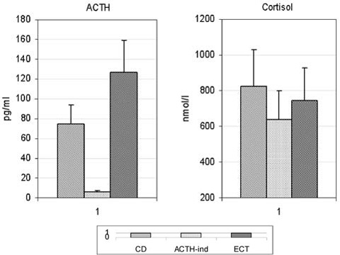
Figure 1. Mean daily concentrations of ACTH and cortisol in patients with hypercortisolism (median+3rd quartile). To convert to SI units for ACTH multiply by 0.22.
Patients with CD (Figure 2) showed a significant rise in ACTH concentration following DM administration: it was 3-9 times the initial ACTH level with a maximum at 15 min after injection (266%; 134-890%), gradually decreasing within the next 90-120 min.
No significant increase of ACTH levels in response to DM was documented in ACTH-ind and ECT patients (Figure 2). The highest ACTH concentration in patients with ECT was 1.5 times (75.0%; 45-117%) the initial level at 15 minutes, whereas in ACTH-ind patients there was practically no response to DM stimulation, [a maximum rise of only 18% (16-50%)] occuring at 30 minutes.
The response of adrenal steroids and ACTH to DM stimulation was not uniform in all patients even in the same group.
The variable secretory responses to DM injections are depicted in Table 2 and are exemplified below.
(1) Variant 1: DM stimulated ACTH secretion and increased the production of steroids by all three zones of the adrenal gland (6 patients with CD).
(2) Variant 2: DM induced secretion of ACTH and Cort but had no effect on Ald and DHEAS secretion (5 patients with CD and 1 with ECT).
(3) Variant 3: DM induced secretion of ACTH and Cort in association with either Ald or DHEAS; 5 patients with CD, [1 with increased Cort and DHEAS (3a) and 4 (3b) with increased Cort and Ald levels].
(4) Variant 4: Administration of DM resulted in an increased secretion of Cort, Ald, and DHEAS in various combinations without a rise in ACTH:
4a- stimulation of all studied steroids (1 patient with ACTH-ind)
4b- stimulation of Cort + Ald (1 patient with ACTH-ind)
4c- stimulation of Cort + DHEAS (2 patients with ACTH-ind)
4d- stimulation of Cort secretion (1 patient with ACTH-ind)
4e- stimulation of Ald + DHEAS secretion (1 patient with CD and 1 patient with ECT)
4f- stimulation of DHEAS secretion (2 patients with CD, 1 with ECT and 1 with ACTH-ind)
4g- stimulation of Ald secretion (2 patients with ECT).
(5) Variant 5: DM did not stimulate the adrenal cortex (2 patients with CD, 6 patients with ECT and 5 patients with ACTH-ind).
It follows from Table 2 that the majority of CD patients (16 of the 21) responded to the administration of DM by increased secretion of ACTH and one or more steroids of the adrenal cortex. In contrast, the adrenal cortex of patients with ACTH-ind and ECT (10 of the 22) responded to DM without a concomitant rise of ACTH. One patient with ECT was an exception. In this patient a rise in Cort followed ACTH elevation.
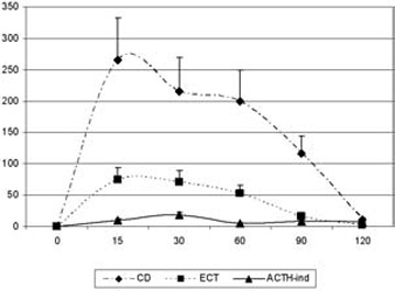
Figure 2. Variations of plasma ACTH level in patients with hypercortisolism in response to desmopressin (median+3rd quartile). Percent (%) rise from baseline
Variant 1. Administration of DM to patients with CD triggered a good ACTH surge at 15 minutes after injection with a 342% rise (134-854%) over the initial level. It should be emphasized that these patients showed the most pronounced response to DM compared with patients of the other groups (Figure 2). An increased level of ACTH in CD patients was associated with a 95.5% (34-180%), 84.5% (45-160%), and 81% (18-110%) rise in Cort, DHEAS, and Ald concentrations, respectively. Maximum values of these steroids were observed within 30-60 min after the rise of plasma ACTH concentration (Table 3 ).
Variant 2. Administration of DM to patients with CD triggered a significant ACTH rise 15 minutes after injection [228.5%; (34-563%)] and in patients with ECT a 75% rise. The Cort value increased by 62% (36-245%) at 15 min and 92% (23-282%) at 60 min, without any change in Ald and DHEAS levels (Table 3 ).
Variant 3. Administration of DM to patients with CD triggered a significant ACTH surge 15 minutes after injection [230% (148-800%)]. The adrenal cortex responded by a rise of Cort and DHEAS (173% and 195%, respectively) or Cort and Ald (135% and 76%, respectively). The concentrations of Ald in the former and DHEAS in the latter case remained virtually unaltered.
Variant 4. Administration of DM resulted in an increased secretion of Cort, Ald, and DHEAS in different combinations without a rise in ACTH. This variant of adrenal response to DM was observed in patients with all forms of hypercortisolism studied. However, it was detected in almost 50% of the subjects with ACTH-ind and ECT and 14% of the patients with CD.
Variant 5. The adrenal cortex in almost half of the patients with ACTH-ind (5 of the 11) and of the ECT patients (6 of the 11) and 3 patients with CD failed to respond to DM administration.
DISCUSSION
The present study has demonstrated marked individual variations of corticosteroid secretion in response to the administration of DM in patients with various forms of hypercortisolism.
The stimulatory action of VP and its analogues on the secretion of pituitary ACTH made it possible to use the DM-test for the differential diagnosis of CD and ECT. Most authors have recorded a rise in plasma ACTH level within 15-30 min after DM injection followed by an increase of Cort 30-45 min later.7 Patients with CD and ECT differ in the degree of such elevation: the former show a 60-100% rise in the level of ACTH, whereas in the latter ACTH concentration increases by less than 30%.8 Our observations are in excellent agreement with the results of other authors. In the present study, there was a 5 to 10-fold rise in the ACTH concentration following a single DM injection in patients with CD compared with a 1.5-fold increase in patients with ECT. No change in the ACTH level was recorded in patients with ACTH-ind.
It is known that steroidogenesis in the adrenal cortex is regulated not only by ACTH but also by a variety of biologically active compounds released into the circulation, such as neuropeptides, neurotransmitters, cytokines, etc. It has been demonstrated in vivo and in vitro that these agents also include VP, which has been shown to stimulate the biosynthesis of Ald in rat adrenal cells9 as well as Ald and Cort in bovine adrenal glands,10 possibly via the activation of V1-receptors. V1-receptors are mainly present in the zona reticularis, with smaller quantities localized in the zona glomerulosa and zona fasciculate.10 Perfusion of isolated canine adrenal glands with a VP-containing solution revealed that cells in the zona fasciculata are more sensitive to the direct action of VP than cells in the zona glomerulosa.6 Also, it is noteworthy that VP stimulates adrenal blood flow and thereby increases Cort secretion.11
The mechanism of enhanced Cort production in patients with ACTH-ind Cushing’s syndrome, when the ACTH production in the adenohypophysis is almost completely suppressed, is not clear. Only recently, adrenal tumours and hyperplastic cortical tissue were shown to contain unusual membrane receptors called “ectopic hormonal receptors”.3 They bind hormonally active compounds and result in Cort biosynthesis, secretion and, possibly, in cell proliferation. The understanding of the exact mechanism by which a biological effect is mediated through the “ectopic receptors” will potentially introduce novel modalities for the conservative therapy of ACTH-ind Cushing’s syndrome and CD. As an example, GIP activation of steroidogenesis can be prevented by GIP blockers and the gonadotropin secretion can be suppressed by long-acting GnRH analogues.
It may be hypothesized that the presence of active “ectopic receptors” in tumours is a general biological phenomenon; in other words, hormone-producing tumours are not autonomous but functionally dependent on a variety of biologically active compounds that exert their influence through the corresponding “ectopic receptors”.
The fact that the administration of exogenous vasopressin to patients with different forms of Cushing’s syndrome stimulates biosynthesis of Cort, Ald, and androgens in the absence of increased production of ACTH12 suggests the occurrence of “ectopic” V1-receptors in human adrenal glands. However, investigations into the synthesis and secretion of individual adrenal steroids are few.
Our study revealed certain unique responses of the adrenal cortex to DM administered to patients with various forms of hypercortisolism.
The overwhelming majority of patients with CD responded to exogenous vasopressin by a rise in ACTH secretion and subsequent increase of plasma Cort concentration (16 out of 21 patients). This observation indicates that the adrenal glands of these patients remain susceptible to the ACTH action and that the function of their pituitary-adrenal axis remains intact. In six patients with CD, ACTH had an effect on all three zones of the adrenal gland, in five patients on two zones (either glomerulosa and fasciculata or reticularis and fasciculata), and in the remaining five on the zone fasciculata alone. In two patients with CD, DM apparently acted directly on the adrenal cortex, stimulating only DHEAS release in two patients and Ald and DHEAS secretion in one patient.
In patients with ECT, secretion of DHEAS and Ald, unlike that of Cort, remained unaltered despite a rise in the ACTH concentration. Nevertheless, the adrenal cortex responded to DM injections by elevated secretion of Cort (one patient), Ald (two patients), DHEAS (one patient) or both DHEAS and Ald (one patient).
The group of patients with ACTH-ind includes six patients with a unique response of the adrenal cortex to DM; there was a rise of Cort (one patient), of DHEAS (one patient), of Cort and Ald (one patient), of Cort and DHEAS (two patients), and of Cort, Ald and DHEAS (one patient). None of the patients with ACTH-ind responded to the administration of DM by an increase of ACTH secretion.
The results obtained in this study suggest expression of DM receptors (“ectopic receptors”, possibly V1 and/or V2-receptors)13 in the three cortical zones of the adrenal gland regardless of the etiology of hypercortisolism. Specifically, the data showing a rise of Cort in five patients, of Ald in six patients, and of DHEAS in nine patients, following DM administration without a rise in ACTH values, favour the presence of V1 and/or V2 receptor in the adrenal cortex. Cort secretion, as a rule, increased in association with elevated plasma ACTH concentration, that is, as a result of stimulation of Cort biosynthesis by ACTH.
It is obvious that the DM test revealed marked heterogeneity in the response of adrenal steroids to DM in all forms of hypercortisolism. This suggests that the development of hypercortisolism may be mediated through diverse mechanisms even within a single form of hypercortisolism.5,14
CONCLUSION
1. The DM test provides a reliable tool for the differential diagnosis of individual variants of hypercortisolism of diverse etiology, such as Cushing’s disease, ectopic ACTH-dependent Cushing’s syndrome, and Cort secreting adrenal tumour.
2. Desmopressin directly stimulates steroidogenesis in the adrenal cortex in certain patients with hypercortisolism, possibly acting via “ectopic receptors”.
REFERENCE
1. Ross NS, 1994 Epidemiology of Cushing’s syndrome and subclinical disease. Endocrinol Metab Clin North Am 23: 539-546.
2. Betragna X, 1994 Proopiomelanocortin-derived peptides. Endocrinol Metab Clin North Am 23: 467-485.
3. LaCroix A, N’Diaye N, Tremblay J, et al, 2001 Ectopic and abnormal hormone receptors in adrenal Cushing’s syndrome. Endocrin Rev 22: 75-110.
4. Goncharov N, Kolesnikova G, Vorontsov V, et al, 1996 Use of radioimmunoassay methods in the characterization of steroidogenesis in hormone-producing adrenal cortex tumors. Proc. 6th Symp. On the Analysis of Steroids. Szeged, Hungary; pp, 407-425.
5. Mune T, Murase H, Yamakita N, et al, 2002 Eutopic overexpression of vasopressin v1a receptor in adrenocorticotropin-independent macromolecular adrenal hyperplasia. J Clin Endocrin Metab 87: 5706-5713.
6. Schneider EG, 1988 Effect of vasopressin on adrenal steroidogenesis. Am J Physiol 255: 806-811.
7. Losa M, Mortini P, Dylgjeri S, et al, 2001 Desmopressin stimulation test before and after pituitary surgery in patients with Cushing’s disease. Clin Endocrin (Oxf) 55: 61-68.
8. Sakai Y, Horiba N, Tozawa F, et al, 1997 Desmopressin stimulation test for diagnosis of ACTH-dependent Cushing’s syndrome. Endocr J 44: 687-695.
9. Gallo-Payet N, Guillon G, 1998 Regulation of adrenocortical function by vasopressin. Horm Metab Res 30: 360-367.
10. Bird IM, Nicol M, Williams BC, et al, 1990 Vasopressin stimulates cortisol and phosphoinositide catabolism in cultured bovine adrenal fasciculata/reticularis cells. J Mol Endocrinol 5: 109-116.
11. Nussdorfer GG, 1996 Paracrine control of adrenal cortical function by medullary chromaffin cells. Pharmacol Rev 48: 495-530.
12. LaCroix A, Tremblay J, Touyz RM, et al, 1997 Propanol Therapy for ectopic beta-adrenergic receptors in adrenal Cushing’s syndrome. J Clin Endocrinol Metab 82: 2414-2422.
13. Tsagarakis S, Tsigos C, Vasiliou V, et al, 2002 The desmopressin and combined CRH-desmopressin tests in the different diagnosis of ACTH-dependent Cushing’s syndrome: constraints imposed by the expression of V2 vasopressin receptors in tumors with ectopic ACTH secretion. J Clin Endocrinol Metab 87: 1646-1653.
14. Cavagnini F, Pecori Giraldi F, 2001 Epidemiology and follow-up of Cushing’s disease. Ann Endocrinol (Paris) 62: 168-172.