HORMONES 2012, 11(1): 77–85
DOI: 10.1007/BF03401540
Ekaterini Koiou,1 Konstantinos Tziomalos,2 Ilias Katsikis,1 Konstantinos Dinas,1 Elena A. Tsourdi,1 Eleni A. Kandaraki,1 Dimitrios Delkos,1 Efstathios Papadakis,1 Dimitrios Panidis1
1Division of Endocrinology and Human Reproduction, Second Department of Obstetrics and Gynecology, Hippokration Hospital, 2First Propedeutic Department of Internal Medicine, AHEPA Hospital, Aristotle University of Thessaloniki, Thessaloniki, Greece
Address for correspondence:
Konstantinos Tziomalos, MD, PhD, First Propedeutic Department of Internal Medicine, AHEPA Hospital, 1 Stilponos Kyriakidi Str.,546 36, Thessaloniki, Greece, Tel.: +30 2310994621, Fax: + 30 2310274434, e-mail: ktziomalos@yahoo.com
Received 25-07-11, Revised 07-10-11, Accepted 12-01-11
Abstract
OBJECTIVE: We aimed to assess plasma Von Willebrand factor (vWF) levels in women with polycystic ovary syndrome (PCOS) and to compare these levels among the different PCOS phenotypes. DESIGN: We studied 140 women with PCOS and 40 age and body mass index (BMI)- matched healthy women (control group). RESULTS: Plasma vWF antigen levels were higher in women with PCOS than in controls (p=0.017). Plasma vWF antigen levels were also higher in patients with phenotypes 1 [i.e. with anovulation (ANOV), biochemical hyperandrogenemia or clinical manifestations of hyperandrogenemia (HA) and polycystic ovaries (PCO)] and 2 (i.e. with ANOV and HA but without PCO) than in controls (p=0.017). In contrast, plasma vWF antigen levels did not differ between controls and patients with phenotypes 3 (i.e. with HA and PCO but without ANOV) and 4 (i.e. with ANOV and PCO but without HA) or between patients with phenotypes 1 and 2 and patients with phenotypes 3 and 4. When overweight/obese and normal weight subjects were analyzed separately, plasma vWF antigen levels did not differ between patients with PCOS (regardless of phenotype) and controls. CONCLUSIONS: Plasma vWF levels are elevated in women with PCOS. This increase appears to be more pronounced in women with phenotypes 1 and 2 of PCOS. Given the association between vWF levels and increased incidence of cardiovascular events, the evaluation of vWF levels in women with PCOS might be helpful for cardiovascular risk stratification, but prospective studies are needed to support this hypothesis.
Key words: Cardiovascular risk, Insulin resistance, Obesity, Polycystic ovary syndrome, Polycystic ovary syndrome phenotypes, Von Willebrand factor
INTRODUCTION
Polycystic ovary syndrome (PCOS) is one of the commonest endocrine disorders in women of reproductive age.1 Obesity and insulin resistance (IR) are frequently present in patients with PCOS.1 Both obesity and IR are associated with increased cardiovascular risk2,3 and accumulating data suggest that women with PCOS have a higher incidence of cardiovascular events than age-matched controls.4 Women with the “classic” PCOS phenotype introduced by the National Institute of Health criteria in 19905 have more adverse metabolic characteristics than women with the additional PCOS phenotypes proposed in 2003 by the European Society for Human Reproduction and Embryology and the American Society for Reproductive Medicine-Sponsored Consensus Group.6,7 However, it is unclear whether cardiovascular risk differs between women with different PCOS phenotypes.4
Von Willebrand factor (vWF) plays an important role in the pathogenesis of atherothrombosis.8 First, vWF mediates platelet adhesionto the injured endothelium as well as platelet aggregation, which are the first steps in thrombus formation.8 Second, as vWF is produced almost exclusively in endothelial cells, elevated plasma vWF levels are a marker of endothelial dysfunction;9,10 in turn, endothelial dysfunction increases the risk for cardiovascular events.11 Accordingly, several prospective studies have shown that elevated plasma vWF levels are independently associated with higher incidence of cardiovascular events.12 Interestingly, a recent analysis of the Framingham Offspring Study showed that the association between elevated vWF levels and cardiovascular risk is stronger in patients with IR than in those without IR.13 Given the high prevalence of IR in PCOS,1 this observation is particularly pertinent in women with PCOS.
Both IR and obesity are considered major determinants of plasma vWF levels and both are associated with elevated vWF levels.14,15 Therefore, it is reasonable to assume that plasma vWF levels are elevated in PCOS, i.e. a syndrome characterized by a high prevalence of both IR and obesity.1 However, none of the several studies that evaluated circulating vWF levels in PCOS showed a significant difference in these levels between women with PCOS and control women.16-20 In addition, there are no studies that evaluated vWF levels in the different PCOS phenotypes. Accordingly, the aim of the present study was to evaluate plasma vWF levels in a larger population of women with PCOS, to assess these levels in overweight/obese and normal weight subjects separately and to compare them among the four PCOS phenotypes. We also aimed to determine whether the anthropometric, metabolic and endocrine characteristics of women with PCOS correlate with plasma vWF levels, with particular emphasis on obesity, IR and hyperandrogenemia.
SUBJECTS AND METHODOLOGY
Patients
We studied 140 women with PCOS and 40 age and body mass index (BMI)-matched healthy women (control group). All women with PCOS were outpatients at the Gynecological Endocrinology Infirmary of the Second Department of Obstetrics and Gynecology, Aristotle University of Thessaloniki, Greece. Women of the control group were healthy volunteers with normal ovulating cycles (28±2 days, blood progesterone levels >10 ng/ml in two consecutive cycles), no signs of hyperandrogenism and normal sonographic appearance of the ovaries. Even though we tried to include more women in the control group, this was not feasible due to the strict inclusion criteria and because most healthy women at this age are unmarried, not contemplating pregnancy and therefore not interested in undergoing hormonal tests.
Diagnosis of PCOS was based on the revised criteria of Rotterdam.6 None of the women studied had galactorrhea or any endocrine or systemic disease that could possibly affect reproductive physiology. No woman reported use during the last semester of any medication that could interfere with the normal function of the hypothalamic-pituitary-gonadal axis. When basic 17α-hydroxyprogesterone (17α-OHP) levels were >1.5ng/ml, the Synacthen test (0.25 mg/1ml; Novartis Pharma S.A., Rueil-Malmaison, France) was performed to rule out congenital adrenal hyperplasia. Other causes of hyperandrogenemia, including prolactinoma, Cushing’s syndrome and androgen-secreting tumors, were also excluded.
Informed consent was obtained from all women and the study was approved by the institutional review board. The study met the requirements of the 1975 Helsinki guidelines.21
Study protocol
In all women, body weight, height and waist circumference (WC) were measured. Body weight was measured with analog scales and in light clothing; height was measured barefoot with a stadiometer. The BMI was calculated by dividing weight (in kg) by height squared (in m) to assess obesity. The WC was defined as the smallest circumference at the level of the umbilicus.
Baseline blood samples were collected between days 3 and 7 of the menstrual cycle in the control group and between 3 to 7 days after a spontaneous bleeding episode in patients with PCOS, after an overnight fast. The circulating levels of follicle-stimulating hormone (FSH), luteinizing hormone (LH), prolactin (PRL), testosterone (T), Δ4-androstenedione (Δ4-A), dehydroepiandrosterone sulfate (DHEA-S), 17α-OHP, sex hormone-binding globulin (SHBG), glucose, insulin, thyroid stimulating hormone (TSH) and free thyroxin (FT4) were measured. Immediately after the baseline blood sampling an oral glucose tolerance test (OGTT) was performed; 75 g of glucose were administered orally and serum glucose levels were determined after 30, 60, 90 and 120 min. On the same day, transvaginal ultrasonography was performed and the volume of each ovary and the number of follicles in each ovary were determined.
Patients with PCOS and controls were divided according to BMI into normal weight (BMI <25 kg/m2; n=70 patients with PCOS and n=20 controls) and overweight/obese (ΒΜΙ >25 kg/m2; n=70 patients with PCOS and n=20 controls) (Figure 1).
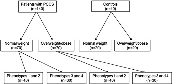
Figure 1. Description of study groups.
Normal weight and overweight/obese patients with PCOS were further subdivided into patients with phenotypes 1 and 2 (n=40 and n=40, respectively) and patients with the additional PCOS phenotypes introduced by the 2003 criteria (n=30 patients with phenotypes 3 and 4 and n=30 patients with phenotypes 3 and 4) (Table 1 and Figure 1).6 Patients with phenotype 1 (“severe PCOS”) had oligo- or anovulation (<8 spontaneous hemorrhagic episodes/yr), biochemical hyperandrogenemia (early follicular phase testosterone >60 ng/dl, corresponding to the mean±2 SD of 200 control subjects measured in our laboratory) or clinical manifestations of hyperandrogenemia (Ferriman-Gallwey score ?8), and polycystic ovaries on ultrasound (?12 follicles with a diameter of 2-9 mm in at least 1 ovary and/or ovarian volume >10cm3) (Table 1). Patients with phenotype 2 had oligo- or anovulation, biochemical hyperandrogenemia or clinical manifestations of hyperandrogenemia and normal sonographic appearance of the ovaries (Table 1). Patients with the additional PCOS phenotypes introduced by the 2003 criteria had biochemical hyperandrogenemia or clinical manifestations of hyperandrogenemia and polycystic ovaries without oligo- or anovulation [phenotype 3 (“ovulatory” PCOS)] or had oligo- or anovulation and polycystic ovaries, without biochemical hyperandrogenemia or clinical manifestations of hyperandrogenemia [phenotype 4 (“mild” PCOS)] (Table 1).
METHODS
Plasma glucose, insulin, FSH, LH, PRL, androgens, 17α-OHP, TSH and FT4 concentrations were measured as previously described.22 Plasma vWF antigen levels were determined by immuno-turbidimetry (Dade Behring vWF Ag® test, Dade Behring, Marburg, Germany). The intra- and inter-assay coefficients of variation were 1.4 and 2.1%, respectively, and the reference range was 50-120%. Free androgen index (FAI) was determined as follows: FAI = T (nmol/l) x 100 / SHBG (nmol/l).23 The homeostasis model assessment of IR (HOMA-IR) index was calculated as follows: HOMA-IR = fasting insulin (mIU/l) x glucose (mg/dl) / 405.24 The quantitative insulin sensitivity check index (QUICKI) was calculated according to the following formula: QUICKI = 1/[log Insulin (mIU/l) + log Glucose (mg/dl)].25
Transvaginal ultrasonography
Transvaginal ultrasonography was performed in all women by an experienced operator. Ovarian volume was calculated as follows: ovarian volume = (π/6) x ovarian length x ovarian height x ovarian width.
Statistical analysis
Frequency matching was used to match women with PCOS and controls for age and BMI. Data analysis was performed with the statistical package SPSS (version 17.0; SPSS Inc., Chicago, IL). Data are reported as mean±SD. Because several tested parameters did not follow normal distribution as assessed with the Kolmogorov-Smirnov test, the Mann-Whitney test was used for comparisons between groups. In addition, because we compared 22 different variables between patients with PCOS and controls, we adjusted the p value for multiple comparisons according to the criteria proposed by Kusuoka and Hoffman;26 accordingly, a p value <0.031 was considered significant. Correlations between vWF levels and other parameters were assessed via Spearman Rank Order correlation. Parameters that were significantly correlated with vWF levels according to Spearman’s correlation were included in a stepwise linear regression analysis model to identify independent correlations with vWF levels.
RESULTS
Characteristics of the total study population are shown in Tables 2 and 3 . The mean age of the women with PCOS (n=140) was 23.9±4.9 years and their mean BMI was 27.2±6.6 kg/m2. The mean age of the controls (n=40) was 23.8±3.3 years and their mean ΒΜΙ was 24.9±3.7 kg/m2. Women with PCOS had lower plasma FSH and SHBG levels and higher plasma T, Δ4-A, DHEA-S, FAI and 17α-OHP levels than controls. In addition, women with PCOS had greater mean ovarian volume and a higher mean number of ovarian follicles than controls. There were no differences in markers of IR (plasma glucose and insulin levels, the glucose/insulin ratio, the area under the OGTT curve and the indices HOMA-IR and QUICKI) between women with PCOS and controls. Plasma vWF antigen levels were higher in women with PCOS compared with controls (57.1±27.7 vs. 45.9±21.9%, respectively; p=0.017; Table 3 ). In addition, patients with phenotypes 1 and 2 of PCOS had higher plasma vWF antigen levels than controls (57.7±28.2 and 45.9±21.9, respectively; p=0.017; Figure 2). In contrast, plasma vWF antigen did not differ between patients with phenotypes 3 and 4 of PCOS and controls (56.3±27.2 and 45.9±21.9, respectively; Figure 2) or between patients with phenotypes 1 and 2 of PCOS and patients with phenotypes 3 and 4 of PCOS (57.7±28.2 and 56.3±27.2, respectively; Figure 2).
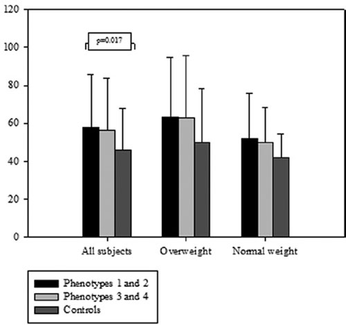
Figure 2. Plasma Von Willebrand factor (vWF) antigen levels according to the body mass index and the phenotype of polycystic ovary syndrome.
Characteristics of overweight/obese women with PCOS and controls are shown in Tables 2 and 3 . Overweight/obese women with PCOS had greater BMI than controls. Plasma androgen levels were also higher in the former. Markers of IR did not differ between overweight/obese women with PCOS and controls. Overall, plasma vWF antigen levels did not differ between overweight/obese women with PCOS and controls. In addition, plasma vWF antigen levels did not differ significantly between overweight/obese patients with phenotypes 1 and 2 of PCOS, overweight/obese patients with phenotypes 3 and 4 of PCOS and overweight/obese controls (63.5±31.1, 62.9±32.8 and 49.9±28.2, respectively; Figure 2).
Characteristics of normal weight women with PCOS and controls are shown in Tables 2 and 3 . Again, normal weight women with PCOS had higher plasma androgen levels than controls. The area under the OGTT curve was greater in women with PCOS and was the only marker of IR that differed significantly between the two groups. Plasma vWF antigen levels did not differ between normal weight women with PCOS and controls. In addition, plasma vWF antigen levels did not differ significantly between normal weight patients with phenotypes 1 and 2 of PCOS, normal weight patients with phenotypes 3 and 4 of PCOS and normal weight controls (51.8±23.9, 49.7±18.4 and 41.8±12.5, respectively; Figure 2).
Overweight/obese women with PCOS had higher plasma vWF antigen levels than normal weight women with PCOS (63.2±31.6 and 50.9±21.6, respectively; p=0.029).
In the total population, in univariate analysis, plasma vWF antigen levels correlated with age (r=0.175, p=0.019), the FAI (r=0.152, p=0.041) and markers of obesity [BMI (r=0.237, p=0.001) and WC (r=0.170, p=0.022)] and IR [plasma glucose (r=0.179, p=0.016) and insulin levels (r=0.216, p=0.004), glucose/insulin (r= -0.183, p=0.014), HOMA-IR (r=0.242, p=0.001) and QUICKI (r= -0.242, p=0.001)]. In multivariate analysis, plasma vWF levels independently correlated with age, BMI, WC, plasma glucose levels and QUICKI (p=0.004, p=0.003, p=0.021, p=0.037 and p=0.043, respectively).
In women with PCOS, in univariate analysis, plasma vWF antigen levels correlated with age (r=0.256, p=0.002) and markers of obesity [BMI (r=0.252, p=0.003) and WC (r=0.196, p=0.020)]. In multivariate analysis, plasma vWF levels independently correlated with age and BMI (p=0.002 for both correlations).
In controls, in univariate analysis, plasma vWF antigen levels correlated with plasma PRL levels (r=0.316, p=0.047) and with markers of IR [plasma glucose (r=0.420, p=0.007) and insulin levels (r=0.511, p=0.001), glucose/insulin (r=-0.434, p=0.005), HOMA-IR (r=0.544, p<0.001) and QUICKI (r= -0.544, p<0.001)]. In multivariate analysis, plasma vWF levels independently correlated with the QUICKI (p=0.001).
DISCUSSION
We report significantly higher plasma vWF levels in women with PCOS compared with age- and BMI-matched healthy volunteers. This finding is in contrast with previous studies, which did not detect differences in circulating vWF levels between women with PCOS and controls.16-20 However, the present study is the largest that assessed plasma vWF levels in PCOS (n=140) and this allowed us to detect an increase in plasma vWF levels in women with PCOS. Indeed, when we analyzed overweight/obese and normal weight subjects separately, there were no differences in plasma vWF levels between patients and controls in these smaller subgroups (n=70). Accordingly, in the largest study of vWF levels to date, Moran et al., who evaluated a comparable number of overweight/obese women with PCOS (n=80), did not find an increase in vWF levels compared with controls.18 Moreover, in some previous studies, women with PCOS were not age-16,20 and BMI-matched20 with controls. This might have obscured the differences in vWF levels between the two groups because vWF levels correlated with age in the present study and with BMI in both the present and previous studies.18
This is the first study that evaluated circulating vWF levels in the four different phenotypes of PCOS. It is well established that patients with phenotypes 1 and 2 have a more adverse metabolic profile than patients with phenotypes 3 and 4.7 However, plasma vWF levels did not differ between these two groups in our study. Nevertheless, in the total study population, plasma vWF antigen levels were higher in patients with phenotypes 1 and 2 than controls but did not differ between patients with phenotypes 3 and 4 and controls. Therefore, it appears that the increase in plasma vWF antigen levels in phenotypes 1 and 2 is the main driver of the elevated vWF levels in PCOS. These differences however were not observed when overweight/obese and normal weight subjects were analyzed separately. Clearly, more studies are needed to confirm that phenotypes 1 and 2 are associated with a greater increase in vWF levels than phenotypes 3 and 4.
Insulin resistance is a risk factor for endothelial dysfunction and results in elevated vWF levels.15 However, in our study, plasma vWF levels did not correlate with markers of IR in women with PCOS and markers of IR did not differ between women with PCOS and controls. In addition, in previous studies in both non-obese17,19 and obese women with PCOS,18 vWF levels did not correlate with indices of IR. Moreover, treatment with metformin of overweight/obese women with PCOS did not affect vWF levels despite an improvement in IR.27 More studies are therefore required to determine the contribution of IR to the increased vWF levels in PCOS.
In our study, plasma vWF levels did not correlate with circulating androgens in women with PCOS and this is in agreement with previous studies in both non-obese19 and obese women with PCOS.18 In addition, plasma vWF levels did not differ between patients with phenotypes 1 and 2 of PCOS and patients with phenotypes 3 and 4 despite the more marked hyperandrogenemia in the former. Moreover, treatment of overweight/obese women with PCOS with oral contraceptives (ethinyl estradiol plus cyproterone acetate) had no effect on vWF levels despite a reduction in plasma androgen levels.27 Finally, administration of testosterone to healthy females did not affect plasma vWF levels.28 Circulating androgens therefore do not appear to affect vWF levels in PCOS.
Plasma vWF levels were rather low in the controls in the present study. It is possible that the young age of our population played a role in these low levels, since age directly correlated with vWF levels in our study. In addition, samples were stored deep frozen at -70oC and this might have also resulted in low vWF levels. Indeed, previous studies reported that cold-temperature storage of blood leads to significant loss of vWF.29 However, all samples from both patients and controls were thawed at the same time and tested in the same run. We thus believe that our findings regarding the comparison of plasma vWF levels between patients and controls are valid.
In conclusion, our findings suggest that plasma vWF levels are elevated in women with PCOS. This increase appears to be more pronounced in women with phenotypes 1 and 2 of PCOS. Insulin resistance potentially mediates this increase in vWF levels, whereas hyperandrogenemia does not appear to play a role. Given the association between vWF levels and increased incidence of cardiovascular events,12 the evaluation of vWF levels in women with PCOS might be helpful for cardiovascular risk stratification, but prospective studies are needed to support this hypothesis.
REFERENCES
1.Norman RJ, Dewailly D, Legro RS, Hickey TE, 2007 Polycystic ovary syndrome. Lancet 370: 685-697.
2.Poirier P, Giles TD, Bray GA, et al, American Heart Association; Obesity Committee of the Council on Nutrition, Physical Activity, and Metabolism, 2006 Obesity and cardiovascular disease: pathophysiology, evaluation, and effect of weight loss: an update of the 1997 American Heart Association Scientific Statement on Obesity and Heart Disease from the Obesity Committee of the Council on Nutrition, Physical Activity, and Metabolism. Circulation 113: 898-918.
3.Nigro J, Osman N, Dart AM, Little PJ, 2006 Insulin resistance and atherosclerosis. Endocr Rev 27: 242-259.
4.Wild RA, Carmina E, Diamanti-Kandarakis E, et al, 2010 Assessment of cardiovascular risk and prevention of cardiovascular disease in women with the polycystic ovary syndrome: a consensus statement by the Androgen Excess and Polycystic Ovary Syndrome (AE-PCOS) Society. J Clin Endocrinol Metab 95: 2038-2049.
5.Zawadski JK, Dunaif A 1992 Diagnostic criteria for polycystic ovary syndrome: towards a rational approach. In: Dunaif A, Givens JR, Haseltine FP, Merriam GE, Hershman SM (eds) Polycystic ovary syndrome. Current issues in Endocrinology and Metabolism. Blackwell, Boston; pp, 377-384.
6.Rotterdam ESHRE/ASRM-Sponsored PCOS Consensus Workshop Group, 2004 Revised 2003 consensus on diagnostic criteria and long-term health risks related to polycystic ovary syndrome (PCOS). Fertil Steril 81: 19-25.
7.Diamanti-Kandarakis E, Panidis D, 2007 Unravelling the phenotypic map of polycystic ovary syndrome (PCOS): a prospective study of 634 women with PCOS. Clin Endocrinol (Oxf) 67: 735-742.
8.Spiel AO, Gilbert JC, Jilma B, 2008 von Willebrand factor in cardiovascular disease: focus on acute coronary syndromes. Circulation 117: 1449-1459.
9.Deanfield JE, Halcox JP, Rabelink TJ, 2007 Endothelial function and dysfunction: testing and clinical relevance. Circulation 115: 1285-1295.
10.Barac A, Campia U, Panza JA, 2007 Methods for evaluating endothelial function in humans. Hypertension 49: 748-760.
11.Kullo IJ, Malik AR, 2007 Arterial ultrasonography and tonometry as adjuncts to cardiovascular risk stratification. J Am Coll Cardiol 49: 1413-1426.
12.Whincup PH, Danesh J, Walker M, et al, 2002 von Willebrand factor and coronary heart disease: prospective study and meta-analysis. Eur Heart J 23: 1764-1770.
13.Frankel DS, Meigs JB, Massaro JM, et al, 2008 Von Willebrand factor, type 2 diabetes mellitus, and risk of cardiovascular disease: the Framingham Offspring Study. Circulation 118: 2533-2539.
14.Gomez-Ambrosi J, Salvador J, Silva C, et al, 2006 Increased cardiovascular risk markers in obesity are associated with body adiposity: role of leptin. Thromb Haemost 95: 991-996.
15.Kim JA, Montagnani M, Koh KK, Quon MJ, 2006 Reciprocal relationships between insulin resistance and endothelial dysfunction: molecular and pathophysiological mechanisms. Circulation 113: 1888-1904.
16.Kelly CJ, Lyall H, Petrie JR, et al, 2002 A specific elevation in tissue plasminogen activator antigen in women with polycystic ovarian syndrome. J Clin Endocrinol Metab 87: 3287-3290.
17.Kuscu NK, Var A, 2009 Oxidative stress but not endothelial dysfunction exists in non-obese, young group of patients with polycystic ovary syndrome. Acta Obstet Gynecol Scand 88: 612-617.
18.Moran LJ, Hutchison SK, Meyer C, Zoungas S, Teede HJ, 2009 A comprehensive assessment of endothelial function in overweight women with and without polycystic ovary syndrome. Clin Sci (Lond) 116: 761-770.
19.Yildiz BO, Bozdag G, Harmanci A, et al, 2010 Increased circulating soluble P-selectin in polycystic ovary syndrome. Fertil Steril 93: 2311-2315.
20.Manneras-Holm L, Baghaei F, Holm G, et al, 2011 Coagulation and fibrinolytic disturbances in women with polycystic ovary syndrome. J Clin Endocrinol Metab 96: 1068-1076.
21.World Medical Association Declaration of Helsinki. Ethical principles for medical research involving human subjects. (available at http://www.wma.net/en/30publications/10policies/b3/17c.pdf) (accessed 30/5/2011).
22.Piouka A, Farmakiotis D, Katsikis I, Macut D, Gerou S, Panidis D, 2009 Anti-Mullerian hormone levels reflect severity of PCOS but are negatively influenced by obesity: relationship with increased luteinizing hormone levels. Am J Physiol Endocrinol Metab 296: E238-243.
23.Carter GD, Holland SM, Alaghband-Zadeh J, Rayman G, Dorrington-Ward P, Wise PH, 1983 Investigation of hirsutism: testosterone is not enough. Ann Clin Biochem 20: 262-263.
24.Matthews D, Hosker J, Rudenski A, Naylor B, Treacher D, Turner R, 1985 Homeostasis model assessment: insulin resistance and beta-cell function from fasting plasma glucose and insulin concentrations in man. Diabetologia 28: 12-19.
25.Katz A, Nambi SS, Mather K, et al, 2000 Quantitative insulin sensitivity check index: a simple, accurate method for assessing insulin sensitivity in humans. J Clin Endocrinol Metab 85: 2402-2410.
26.Kusuoka H, Hoffman JI, 2002 Advice on statistical analysis for Circulation Research. Circ Res 91: 662-671.
27.Teede HJ, Meyer C, Hutchison SK, Zoungas S, McGrath BP, Moran LJ, 2010 Endothelial function and insulin resistance in polycystic ovary syndrome: the effects of medical therapy. Fertil Steril 93: 184-191.
28.Gooren LJ, Giltay EJ, 2008 Review of studies of androgen treatment of female-to-male transsexuals: effects and risks of administration of androgens to females. J Sex Med 5: 765-776.29. Favaloro EJ, 2006 Effect of overnight 4 degrees C storage of whole blood on von Willebrand factor. Transfusion 46: 1057-1059.