HORMONES 2010, 9(2):145-150
DOI: 10.14310/horm.2002.1264
Address for correspondence:
George Dimitriadis, MD, DPhil, 2nd Department of Internal Medicine, Research Institute and Diabetes Center, Athens University Medical School, “Attikon” University Hospital, 1 Rimini Street, 12462 Haidari, Greece, Tel: +30-210-5832547, Fax: +30-210-5832561, E-mail: gdimi@ath.forthnet.gr and gdimitr@med.uoa.gr
Received 10-11-09, Revised 15-01-10, Accepted 10-02-10
Abstract
OBJECTIVE: This study was undertaken to assess the accuracy of GlucoDay® – a portable detector of subcutaneous glucose – by comparing the results to those obtained by Biostator® an established and reliable method for continuous glucose measurement in whole blood. DESIGN: Subjects with type 1 diabetes (n:6), subjects with type 2 diabetes (n:6), and six healthy controls were studied for 24 hours; they consumed three main meals. The GlucoDay® was connected to the subjects by inserting a microfibre probe into the periumbilical subcutaneous area, whilst the Biostator® was inserted by a double-lumen catheter into an antecubital vein. A third catheter was inserted into a separate vein for blood withdrawal to measure glucose by the hexokinase method. RESULTS: The three methods (GlucoDay®-Biostator®-hexokinase) were equally accurate in measuring glucose levels (p=0.233, Kruskall-Wallis test). The glucose measurements performed with GlucoDay® and Biostator® were significantly correlated with those performed with hexokinase (p<0.001, r2=66.65% and p<0.001, r2=64.4%, respectively, using simple regression analysis). CONCLUSIONS: Measurements of glucose fluctuations in the subcutaneous tissue with the GlucoDay® were close to those in blood determined by the Biostator®. GlucoDay® is therefore a reliable method for continuous glucose monitoring and may prove useful for optimizating treatment in patients with type 1 or type 2 diabetes.
Key words: Artificial Pancreas, Diabetes, GlucoDay®, Glucose monitoring, Subcutaneous tissue
INTRODUCTION
It is well established that strict metabolic control protects against the development of chronic complications of diabetes.1,2 Measurements of blood glucose fluctuations are essential for optimization of treatment, but the available methods have limitations (inconvenience and discomfort of finger-pricking, high cost of strips, puncture of veins), which make them unsuitable for frequent estimations of glucose levels. Continuous estimation of blood glucose levels can be performed with the Artificial Endocrine Pancreas (Biostator®),3-5 but this requires an intravenous line.
Changes in glucose levels in the subcutaneous tissue are similar to those in blood.6-9 Observations showing that changes in glucose levels in the subcutaneous tissue are similar to those in blood6-9 opened up the way for the development of portable devices which provide continuous measurement of glucose in the subcutaneous tissue.10-14 Over the last few years, this method has attracted wide clinical interest because patients with diabetes can check their metabolic status on a 24-hour basis, including rarely monitored time periods such as postprandial and nocturnal.15,16
Τhe present study was undertaken to assess the results of glucose monitoring using the GlucoDay® – a new portable detector of subcutaneous glucose17-19 – by comparing them to those obtained by Biostator®, an established method for continuous intravenous glucose monitoring.3-5 As a reference method for both devices, we used glucose measurements in venous blood with hexokinase. These experiments were carried out in subjects with type 1 and type 2 diabetes and in healthy subjects over a 24 hour period.
MATERIAL AND METHODS
We studied 6 subjects with type 1 diabetes (age 30±4years, BMI 23±1kg/m2, HbA1c 6.5±0.6%), 6 subjects with type 2 diabetes (age 59±3years, BMI 27±1kg/m2, HbA1c 6±0.4%) and 6 healthy subjects (age 43±6years, BMI 26±1kg/m2). This study was approved by the hospital ethics committee and subjects gave their written informed consent.
On the day of the study, the subjects were admitted to the Artificial Pancreas Unit of our Department at 07:00 and remained supine for 24 hours. During this time, the patients consumed three main meals (breakfast at 09:00, lunch at 14:00 and dinner at 19:00).
Since the objective of the study was to compare glucose levels measured with the three methods, no attention was paid to the optimization of metabolic control and the diabetic subjects followed their usual treatment schedule (diet, insulin injections or oral anti-diabetic agents).
Three catheters were inserted in each patient: one subcutaneously for connecting the GlucoDay®, one in an antecubital vein for connecting the Biostator® and one for drawing blood to measure glucose with hexokinase.
GlucoDay®
The technical characteristics of GlucoDay® have been previously described.17-19 The microfibre was inserted subcutaneously in the periumbilical region by using an 18-gauge Teflon catheter as a guide, and was then connected to the portable unit worn by the subjects with a belt. It was perfused with Dulbecco’s buffer containing sodium-benzoate as preservative at a rate of 10μl/min. The system makes a glucose measurement every second and stores an average value every 3 minutes, for a total of 480 measurements per day; glucose is measured by glucose oxidase.17-19 The GlucoDay® was calibrated by using a glucose level in venous blood 120 min after the insertion of the microfibre.
In one patient, the GlucoDay® was interrupted after a few hours due to breakage of microfibre and malfunctioning of the device and therefore the number of subjects who completed the study was 17.
Endocrine Artificial Pancreas (Biostator®)
The Biostator® performs blood glucose measurements 6 times per minute (8640 times per 24 hours) via the glucose oxidase method. It was connected to the patients by inserting a double-lumen 18-gauge catheter into an antecubital vein, as previously described.5
Hexokinase
A third catheter was inserted into a separate vein for measurements of glucose in blood samples by hexokinase; this was used as a reference method for both devices. Eight blood samples were withdrawn during the experiment as follows: 120min after the microfibre implantation (this value was used for the calibration of the GlucoDay®), before lunch, 60min after lunch, 120min after lunch, before dinner, 60 min after dinner, 120 min after dinner and immediately before the end of the experiment (at 08:00 the next morning). The specimens were stored in fluoride tubes for glucose determination.
Statistical analysis
The Kolmogorov-Smirnov test was used to examine the normality of data. The Kruskall-Wallis test was performed to test the “null hypothesis” that the medians within each the 3 methods are the same. Correlations were calculated according to the Spearman Rank Correlation coefficient.
We performed simple regression analysis to estimate the slopes of the regression lines. A comparison of alternative models based on the highest r2 value was made in order to find the best fitted between several curvilinear models (i.e. multiplicative, exponential, double reciprocal, logarithmic-X, s-curve, square root-Y, linear, etc.).
Specifically, by performing simple regression analysis to describe the relationship between hexokinase and GlucoDay® and applying the comparison of alternative models, the “double reciprocal model” yielded the highest r2 value. By applying simple regression analysis to describe the relationship between Biostator® and hexokinase and by performing the comparison of alternative models, the “multiplicative model” yielded the highest r2 value. The three methods were then compared with “Multiple Box and Whisker plots”.
Elaboration of data was accomplished by the Stat-graphics Statistical Package (Manugistics, Inc., Rockville, MD). A “p” value less than 5% was considered statistically significant.
RESULTS
No adverse events were observed at the site of GlucoDay® implantation. The device was well tolerated by all patients, even during the overnight recording.
We examined the correlations between each pair of methods (GlucoDay® vs Hexokinase and Biostator® vs Hexokinase). It turned out that GlucoDay® and hexokinase were significantly correlated (r=0.772, p<0.001). Similar results were obtained with Biostator® and hexokinase (r=0.775, p<0.001).
We then compared the median values of the three methods (GlucoDay®-Biostator®-hexokinase) and we found no statistically significant difference amongst the median values (p-value=0.233).
Performing simple regression analysis to describe the relationship between hexokinase and GlucoDay®, the equation of the fitted model was: Glucosehexokinase=1/(0.002+0.863/GlucoseGlucoDay®), r=0.82, r2=66.65%, p<0.001 (Figure 1).
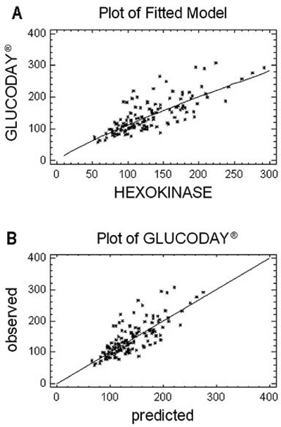
Figure 1. (A) Simple regression analysis of glucose levels (mg/dl)* measured with the GlucoDay® and hexokinase [Y=1/(0.002+0.863/X), r=0.82, r2 =64.36%, p<0.001]. (B) Comparison of glucose levels measured with GlucoDay® and those predicted by the “double reciprocal model”.
* To convert to SI units multiply by 7.175
Applying simple regression analysis to describe the relationship between Biostator® and hexokinase, the equation of the fitted model was: Glucosehexokinase=(2.032)x(GlucoseBiostator®0.846), r=0.80, r2=64.36%, p<0.001 (Figure 2).
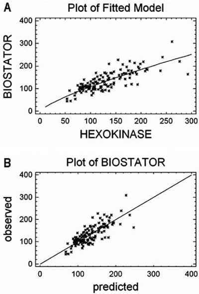
Figure 2. (A) Simple regression analysis of glucose levels (mg/dl)* measured with Biostator® and hexokinase (Y=2.032*X0.846, r=0.80, r2 =64.36%, p<0.001). (B) Comparison of glucose levels measured with the Biostator® and those predicted by the “multiplicative model”.
* To convert to SI units multiply by 7.175
The three methods were then compared with “Multiple Box and Whisker plots”; the width of the boxes representing the interquartile range (Q3 – Q1) was quite similar in all three methods (Figure 3).
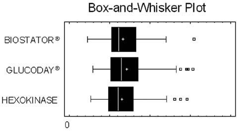
Figure 3. Multiple “Box and Whisker plots” comparing the three methods. The width of the boxes representing the interquartile range (Q3 – Q1) is quite similar in all three compared methods.
DISCUSSION
Adequate monitoring of blood glucose is fundamental for good metabolic control in subjects with type 1 or type 2 diabetes.1,2 For example, information as regards glucose fluctuations in the postprandial period or during sleep is necessary for the improvement of glycaemic control and the prevention of hypoglycaemia. However, self-monitoring of blood glucose cannot provide sufficient information to detect daily variations in glucose profiles,even when performed several times a day.
The possibility of continuous glucose monitoring in diabetic subjects has generated wide clinical interest and has been made possible by the use of the Biostator®.3,4 Indeed, in our study, the Biostator® was accurate in measuring glucose levels, the p-value (with respect to the F distribution test) being less than 0.001, suggesting that there was a significant relationship between glucose levels determined in blood by hexokinase and by the Biostator® at the 99% confidence level (Figure 2). The latter device, however, has serious limitations that restrict its clinical application: it requires an intravenous line and, because of its size, it hampers mobility.
In the last few years, a number of reports have suggested that the changes of glucose levels in the subcutaneous tissue are close to those in peripheral circulation; these observations opened up the way for the development of sensors for continuous subcutaneous glucose monitoring.10-14
In the present study, we assessed the accuracy of the GlucoDay®, a portable detector of glucose in the subcutaneous tissue,17-19 by comparing the results to those obtained by Biostator®, the standard method for continuous glucose determination in blood.3,4 As a reference method for both devices, we used glucose measurements in blood by hexokinase. In a previous study in healthy and diabetic subjects monitored for 24 hours, the GlucoDay® was shown to be reliable and provided measurements of glucose levels that closely agreed with those made in venous blood.19
In our study, the concentrations of glucose in the subcutaneous tissue measured by GlucoDay® were in close agreement with those measured in blood with hexokinase over a wide range of values: the p-value (with respect to the F distribution test) was less than 0.001, suggesting that there was a significant relationship between the two methods at the 99% confidence level (Figure 1). The correlation of GlucoDay® with hexokinase was actually slightly stronger than that of the Biostator® by hexokinase (Figure 2). Since the Biostator® is an established method for continuous glucose monitoring,4 the comparison suggests that the GlucoDay® is a reliable and accurate device for this purpose.
GlucoDay® measures glucose in the interstitial fluid and not in blood. Under physiological conditions, there is a free and rapid exchange of glucose molecules between plasma and interstitial fluid. Therefore, changes in blood glucose and interstitial fluid glucose are expected to correlate well. Nevertheless, changes of glucose concentrations in interstitial fluid do not occur at the same time as those in blood; they lag behind, especially when glycaemia is changing rapidly, for example, after a meal.20,21 This time lag can explain the variation that is apparent between the GlucoDay® and hexokinase measurements as well as the interesting result of non-linearity between the blood and the subcutaneous fluid glucose values (Figure 1).
Despite this limitation, this study compares for the first time results obtained by GlucoDay® with those of Biostator®, showing that continuous glucose measurements in the subcutaneous tissue with GlucoDay® are close to those in blood determined with a standard continuously glucose measuring method (Biostator®).
In conclusion, GlucoDay® is reliable for continuous glucose monitoring in the subcutaneous tissue. This information may prove useful for optimizing treatment in subjects with type 1 or type 2 diabetes mellitus.
ACKNOWLEDGEMENTS
The expert technical help of Annie Triantafylopoulou is gratefully acknowledged. We thank Assoc. Professor M. Sfakianakis PhD for his help with statistical analysis of the data.
REFERENCES
1. DCCT Research Group, 1993 The effect of intensive treatment on the development and progression of long-term complications in insulin-dependent diabetes mellitus. N Eng J Med 329: 977-986.
2. UK Prospective Diabetes Study (UKPDS) Group, 1988 Intensive blood-glucose control with sulphonylureas or insulin compared with conventional treatment and risk of complications in patients with type 2 diabetes. Lancet 352: 837-853.
3. Pfeiffer E, Thum C, Clemens A, 1974 The Artificial beta-cell. A continuous control of blood sugar by external regulation of insulin infusion. Horm Metabol Res 6: 339-342.
4. Pfeiffer E, 1987 On the way to the automated (blood) glucose regulation in diabetes: the dark past, the grey present and the rosy future. Diabetologia 30: 51-65
5. Dimitriadis G, Hatziagelaki E, Ladas S, Linos A, Hillebrand I, Raptis SA, 1988 Effects of prolonged administration of two new a-glucosidase inhibitors on blood glucose control, insulin requirements and breath hydrogen excretion in patients with insulin-dependent diabetes mellitus. Eur J Clin Invest 18: 33-38.
6. Rebrin K, Steil GM, van Antwerp WP, Mastrototaro JJ, 1999 Subcutaneous glucose predicts plasma glucose independent of insulin: implications for continuous monitoring. Am J Physiol 277: E561-E571.
7. Sternberg F, Meyerhoff C, Mennel FJ, Bischof F, Pfeiffer EF, 1995 Subcutaneous Glucose Concentration in humans: real estimation and continuous monitoring. Diabetes Care 18: 1266-1269.
8. Jensen BM, Bjerring P, Christiansen JS, Orskov H, 1995 Glucose content in human skin: relationship with blood glucose levels. Scand J Clin Lab Invest 55: 427-432.
9. Bolinder J, Hagstrom-Toft E, Ungerstedt U, Arner P, 1997 Self- monitoring of blood glucose in type 1 diabetic patients: comparison with continuous microdialysis measurements of glucose in subcutaneous adipose tissue during ordinary life conditions. Diabetes Care 20: 64-70.
10. Moscone D, Mascini M, 1992 Microdialysis and glucose biosensor for in vivo monitoring. Ann Biol Clin (Paris) 50: 323-327.
11. Pfeiffer EF, Meyerhoff C, Bischof F, Keck FS, Kerner W, 1993 Online continuous monitoring of subcutaneous tissue glucose is feasible by combining portable glucosensor with microdialysis. Horm Metab Res 25: 121-124.
12. Pickup JC, 1993 Developing glucose sensors for in vivo use. Trends in Biotechnology 11: 285-291.
13. Meyerhoff C, Bischof F, Mennel FJ, Sternberg F, Pfeiffer FF, 1993 Use of microdialysis technique in the monitoring of subcutaneous tissue glucose concentration. Int J Artif Organs 16: 268-275.
14. Aussedat B, Dupire-Angel M, Gifford R, Klein JC, Wilson GS, Reach G, 2000 Interstitial glucose concentration and glycemia: implications for continuous subcutaneous glucose monitoring. Am J Physiol Endocrinol Metab 278: E716-E728.
15. Turner APF, Pickup JC, 1985 Diabetes mellitus: biosensors for research and management. Biosensors 1: 85-115.
16. Pickup JC, Alcock SJ, 1991 ‘Clinicians’ requirements for in vivo monitoring. Biosens Bioelectron 6: 639-646.
17. Maran A, Poscia A, 2002 Continuous subcutaneous glucose monitoring: the GlucoDay system. Diabetes Nutr Metab 15: 429-433.
18. Maran A, Crepaldi C, Tiengo A, et al, 2002 Continuous subcutaneous glucose monitoring in diabetic patients: a multicenter analysis. Diabetes Care 25: 347-352.
19. Poscia A, Mascini M, Moscone D, et al, 2003 A microdialysis technique for continuous subcutaneous glucose monitoring in diabetic patients. Biosens Bioelectron 18: 891-908.
20. Battelino T, Bolinder J, 2008 Clinical use of real-time continuous glucose monitoring. Curr Diabetes Rev 4: 218-222.
21. De Block C, Vertommen J, Manuel-y-Keenoy B, Van Gaal L, 2008 Minimally-invasive and non-invasive continuous glucose monitoring systems: indications, advantages, limitations and clinical aspects. Curr Diabetes Rev 4: 159-168.