HORMONES 2015, 14(3): 442–446
DOI: 10.14310/horm.2002.1604
Costas Koufaris,1 Angelos Alexandrou,1 Carolina Sismani,1 Nicos Skordis2
1Department of Cytogenetics and Genomics, Cyprus Institute of Neurology and Genetics, Nicosia, Cyprus
2Division of Pediatric Endocrinology, Paedi Center for Specialized Pediatrics, Nicosia, Cyprus
Address for correspondence:
Prof. Nicos Skordis, Division of Pediatric Endocrinology, Paedi Center for Specialized Pediatrics, 178 Athalassas Avenue, 2025 Nicosia, Cyprus; E-mail: nicosskordis@paedi.org.cy
Received: 16-02-2015, Accepted:26-05-2015
Abstract
Familial neurohypophyseal diabetes insipidus (FNDI) is a disorder characterized by excess excretion of diluted urine (polyuria) and increased uptake of fluids (polydipsia). The disorder is caused by mutations affecting the AVP-NPII gene, resulting in absent or deficient secretion of the antidiuretic hormone arginine vasopressin (AVP) by the neurohypophysis. In this study we examined a three-generation Cypriot kindred suspected to have FNDI. Direct sequencing analysis of AVP-NPII identified a missense mutation (NM_000490.4:c.61T>C; p.Tyr21His; rs121964893) within the AVP moiety on exon 1 of the gene in all affected family members. So far, only three studies have reported mutations within the AVP moiety of AVP-NPIIas being associated with FNDI, with the vast majority of identified FNDI mutations being located within the signalling peptide or the neurophysis II (NPII) moiety of the gene. The mutation within the AVP moiety identified here had been reported previously in a Turkish kindred with FNDI. Consequently, the findings of this study confirm the causal role of mutations within the AVP moiety in FNDI. Herein we review reported mutations within the AVP moiety of AVP-NPII and their contribution to FNDI.
Key words: Arginine vasopressin, Autosomal dominant, Founder mutation, Mutation, Neurohypophyseal diabetes insipidus, Sanger sequencing
INTRODUCTION
Diabetes insipidus (DI) is a disorder of water balance, characterized by the excretion of copious amounts of urine and the compensatory consumption of very large quantities of fluids, which can be life-threatening if not managed properly. DI is a rare disease with a nonunivocalreported prevalence of 1: 25,000.1 DI can be acquired as a result of hypothalamic injury or can also be the result of mutations affecting the vasopressin-neurophysin II precursor (AVP-NPII) gene, the AVP receptor 2 (AVPR2) or the aquaporin 2 (AQP2) water channel protein.2
Familial neurohypopheseal dabetes insipidus (FNDI) is caused by mutations affecting the AVP-NPII gene resulting in the absence or deficiency of the anti-diuretic hormone (ADH). This hormone acts to regulate the retention of water through increased water reabsorption in the collecting ducts of the nephron.3
The AVP-NPII gene is an autosomal gene located at 20p13 and is composed of three exons and two introns (Figure 1A). The 19 amino acid long sequence of the signal peptide and the 9 amino acid long sequence of the AVP moiety are encoded within the first exon. The 93 amino acid long N-terminal of the neurophysin II (NPII) is coded across all three exons, with exon 2 coding for the conserved core of NPII and the C-terminal encoded within exon 3. The 39 amino acid glycopeptide known as copeptin is also encoded within exon 3.3 The AVP-NPII product, the AVP preprehormone, is produced in the hypothalamus and is targeted to the endoplasmic reticulum (ER) by the signal peptide. Within the ER the signal peptide is removed and the copeptide is glycosylated. After cleavage and processing, the AVP hormone and its protein carrier NPII are transported to the nearby neurophysin where they can be stored and secreted when necessary to act on the kidney.
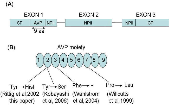
Figure 1. Structure of the AVP-NPII gene and location of FNDI mutations affecting the AVP moiety. (A) Genomic structure of the human AVP-NPII gene. SP signaling peptide, AVP arginine vasopressin, NPII neurophysin II, CP copeptin; (B) mutations within the 9 amino acid AVP moiety reported to be associated with FNDI.
In the majority of cases FNDI is inherited in a completely penetrant, autosomal dominant manner.3 Currently, more than 60 mutations affecting AVP-NPII have been associated with FNDI.1,4 The great majority of FNDI mutations are located within the signal peptide or the NPII moiety of the AVP-NPII gene.3,5-7 However, a few cases have been identified of FNDI caused by mutations within the AVP moiety.8-10
In this study, a three-generation Cypriot family with suspected FNDI was investigated by direct sequence of all three AVP-NPII exons to identify potential pathogenic mutations. A single mutation was identified in affected members of this family within the AVP moiety in exon 2 of the AVP-NPII gene.
MATERIALS AND METHODS
Patients
Five affected members of three-generation FNDI kindred were studied. The pedigree (Figure 2A) is consistent with an autosomal dominant mode of transmission. All members had had polyuria and polydipsia since early childhood but received treatment with desmopressin (DDAVP) relatively late. The proband (patient 2) is an 8-year old boy who had presented with polyuria and polydipsia since infancy. The patient underwent a water deprivation test, which lasted 4 hours and terminated when 25 mcg of DDAVP was administered intranasally that confirmed the diagnosis of central DI. During the test the plasma osmolality increased from 282 to 294 mosm/kg, while the urine osmolality remained low at 150 mosm/kg but increased to 650 mosm/kg following administration of DDAVP. Patient 4, having been symptomatic since infancy, was initially seen at age 17 years. Although the diagnosis was obvious based on the family history, he underwent the water deprivation test which confirmed the diagnosis and an MRI of the pituitary which did not reveal the characteristic bright spot of the posterior pituitary lobe on T1 – weighted images. All patients are in excellent health and doing well on treatment with DDAVP.
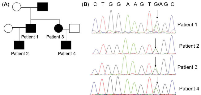
Figure 2. Identification of pathogenic mutation within exon 1 of the AVP-NPII in Cypriot kindred with FNDI. (A) Pedigree of Cypriot family with familial DI.Family members with historical or laboratory evidence for DI are indicated with filled symbols; (B) sequencing chromatogram from a fragment of exon 1 of the AVP-NPII containing identified variant is shown for four examined patients. Arrow shows the location of the missense mutation.
DNA extraction
DNA was extracted using the Qiagen DNA extraction kit (Qiagen Co) according to the manufacturer’s recommendations. The DNA was subsequently quantified using a Nanodrop ND-1000 spectrophotometer.
Sanger sequencing
Primers specific for the three exons of AVP were designed using the Primer3 software.11 The sequence of the primers used for Sanger sequencing of the AVP gene are available upon request. Amplification of the exonic sequences of AVP was performed by PCR using 10ng of genomic DNA and the AmpliTaq polymerase (Invitrogen). PCR products were subjected to gel electrophoresis to ensure the absence of non-specific bands. Purification of PCR products was performed using Exo-SapIT (Affymetrix). Sequencing reactions were performed using Applied Biosystems BigDye terminator v1.1 (Life Technologies) following the manufacturer’s instructions and products were analyzed on an ABI 3130 sequencer. The DNA sequences of the PCR products were mapped to the reference genome using BLAT12 to determine the presence of inherited variants.
RESULTS
Genetic studies
Sanger sequencing of the three exons of the AVP gene identified a single variant that was present in all four affected DI patients, a missense variant it prehormone and position 2 of the mature AVP (NM_000490.4:c.61T>C; p.Tyr21His; rs121964893) (Figure 2B).
DISCUSSION
All the affected kindred discussed herein were found to carry a missense mutation resulting in the substitution of histidine for tyrosine at position 2 of AVP. The same mutation had been previously reported to co-segregate with the disease in a three-generation Turkish kindred with FNDI.8 In those patients, the mutation was associated clinically with inability to concentrate urine during fluid deprivation, a greater than 80% deficiency of AVP secretion and absence of the posterior pituitary bright spot on magnetic resonance imaging. Christensen et al,7 using in vitro models, showed that this mutation retarded the processing of the AVP-NPII prehormone and its accumulation in the endoplasmic reticulum causing reduced secretion. Consequently, this study confirms the causal role of missense mutations affecting this amino acid in the development of FNDI.
The ability of AVP-NPII mutations to cause autosomal dominant FNDI has been proposed as occurring through two mechanisms.1,3 The first mechanism involves the accumulation of the AVP prehormone in the ER of cells, followed by cell dysfunction and death. Autopsies have found loss of AVP-secreting cells in DI patients.13,14 In a mouse model of FNDI, the presence of a truncated version of vasopressin was associated with progressive loss of AVP-producing neurons.15 In a recent publication, evidence has been reported that in an FNDI mouse model, loss of AVP-secreting neurons occurs due to autophagy induced by ER stress.16 A second mechanism involves the direct interaction between the mutant AVP-NPII and the wildtype precursors which blocks its processing in a dominant negative manner. It has been shown in cell models that mutant AVP precursors can form heterodimers with their wildtype counterparts and block their secretion.17 A study has also reported the development of progressive polyuria in a mouse model of FNDI without the loss of vasopressin neurons.18 It is therefore conceivable that loss of AVP-secreting neurons is a nonessential part of FNDI development.
Autosomal dominant FNDI mutations, whether acting by inducing neuronal loss or through interaction of wildtype-mutant AVP precursors, operate by disrupting the physiological folding and processing of AVP-NPII. The majority of these mutations are located within the signalling peptide or in the NPII moiety. For example, Rittig et al5 examined 17 unrelated kindred with FNDI. The researchers identified 13 novel AVP-NPII mutations of which 2 were missense mutations within the signalling peptide, 8 mutations in the NPII moiety in exon 2 and 3 nonsense mutations in the NPII moiety in exon 3. Similarly, Christensen et al19 screened the AVP-NPII gene for mutations in 15 unrelated kindred. This study found 2 novel mutations in the signalling peptide and 4 novel mutations within the NPII moiety. This pattern of the majority of mutations in FNDI located in the NPII moiety followed by the signalling peptide of the AVP precursor has been consistent in a large number of unrelated FNDI kindred studies since then.20
Nevertheless, studies have found a few cases of autosomal dominant mutations associated with FNDI affecting specifically positions 2 and 3 of AVP (Figure 1B). Rittig et al8 found the same missense mutation identified in the present study in Turkish kindred with FNDI. Wahlstrom et al9 reported a 3-base pair deletion resulting in loss of phenylalanine at position 3 of the AVP hormone in an American FNDI kindred. Kobayashi et al10 reported a missense substation at position 2 of AVP of tyrosine by serine in a Japanese patient, the same position found to be affected by Rittig et al8 and by this study, although involving a distinct amino acid substitution.
Crystallography and biochemical data suggest that the dimerisation of NPII with AVP occurs between the first 3 N-terminal amino acids of the hormone and pockets formed by several amino acid residues of NPII.21 It has been proposed that the rarity of mutations within the AVP moiety associated with FNDI relate to the fact that only the first 3 N-terminal amino acids of AVP are essential for the proper processing and folding of the prehormone.8 Another interesting case involved a missense mutation that predicts for a substitution of leucine for proline at position 7 of AVP.22 However, in this case the mutation was considered to be associated with autosomal recessive FNDI, thus it does not possess dominant negative effects and is probably pathogenic due to the loss of the normal functioning protein.
Given the geographic and historical links between the Cypriots and Turkish and other Middle Eastern populations it is difficult to exclude the likelihood that the identical missense mutation identified in this study to the one reported by Rittig8 can be traced back to the same ancestor, i.e. it concerns a founder effect. A large fraction of hereditary endocrinopathies in Cyprus have been associated with founder effects involving previous migrations to the island, in ancient times of Phoenician populations and more recently of people of Turkish, Arab and other ancestries which have shaped the Cypriot gene pool.23 Identical splice-mutations affecting the 5alpha-steroid reductase type 2 gene (SRD5A2) gene—5aSRD IVS1-2A>G—have been found in Cypriots which are most probably due to a founder effect.24
In conclusion, we report here a Cypriot family with FNDI associated with a mutation within the AVP moiety of the AVP-NPII gene. This offers further confirmation that mutations affecting position 2 of AVP are causally implicated in autosomal dominant FNDI.
CONFLICT OF INTEREST
The authors declare no conflict of interest.
Ethical approval
All procedures performed in studies involving human participants were in accordance with the ethical standards of the institutional and/or national research committee and with the 1964 Declaration of Helsinki and its later amendments or comparable ethical standards.
Informed consent
The informed consent was obtained from all individual participants included in the study.
REFERENCES
1. Di Iorgi N, Napoli F, Allegri AE, et al, 2012 Diabetes insipidus–diagnosis and management. Horm Res Paediatr 77: 69-84.
2. Makaryus AN, McFarlane SI, 2006 Diabetes insipidus: diagnosis and treatment of a complex disease. Cleve Clin J Med 73: 65-71.
3. Christensen JH, Rittig S, 2006 Familial neurohypophyseal diabetes insipidus–an update. Semin Nephrol 26: 209-223.
4. Birkegaard C, Christensen JH, Falorni A, et al, A novel variation in the AVP gene resulting in familial neurohypophyseal diabetes insipidus in a large Italian kindred. Pituitary 16: 152-157.
5. Rittig S, Robertson GL, Siggaard C, et al, 1996 Identification of 13 new mutations in the vasopressin-neurophysin II gene in 17 kindreds with familial autosomal dominant neurohypophyseal diabetes insipidus. Am J Hum Genet 58: 107-117.
6. Skordis N, Patsalis PC, Hettinger JA, et al, 2000 A novel arginine vasopressin-neurophysin II mutation causes autosomal dominant neurohypophyseal diabetes insipidus and morphologic pituitary changes. Horm Res 53: 239-245.
7. Christensen JH, Siggaard C, Corydon TJ, et al, 2004 Six novel mutations in the arginine vasopressin gene in 15 kindreds with autosomal dominant familial neurohypophyseal diabetes insipidus give further insight into the pathogenesis. Eur J Hum Genet 12: 44-51.
8. Rittig S, Siggaard C, Ozata M, et al, 2002 Autosomal dominant neurohypophyseal diabetes insipidus due to substitution of histidine for tyrosine(2) in the vasopressin moiety of the hormone precursor. J Clin Endocrinol Metab 87: 3351-3355.
9. Wahlstrom JT, Fowler MJ, Nicholson WE, Kovacs WJ, 2004 A novel mutation in the preprovasopressin gene identified in a kindred with autosomal dominant neurohypophyseal diabetes insipidus. J Clin Endocrinol Metab 89: 1963-1968.
10. Kobayashi H, Fujisawa I, Ikeda K, et al, 2006 A novel heterozygous missense mutation in the vasopressin moiety is identified in a Japanese person with neurohypophyseal diabetes insipidus. J Endocrinol Invest 29: 252-256.
11. Untergasser A, Cutcutache I, Koressaar T, et al, 2012 Primer3–new capabilities and interfaces. Nucleic Acids Res 40: e115.
12. Kent WJ, 2002 BLAT–the BLAST-like alignment tool. Genome Res 12: 656-664.
13. Braverman LE, Mancini JP, McGoldrick DM, 1965 Hereditary idiopathic diabetes insipidus. A case report with autopsy findings. Ann Intern Med 63: 503-508.
14. Bergeron C, Kovacs K, Ezrin C, Mizzen C, 1991 Hereditary diabetes insipidus: an immunohistochemical study of the hypothalamus and pituitary gland. Acta Neuropathol 81: 345-348.
15. Russell TA, Ito M, Ito M, et al, 2003 A murine model of autosomal dominant neurohypophyseal diabetes insipidus reveals progressive loss ofvasopressin-producing neurons. J Clin Invest 112: 1697-1706.
16. Hagiwara D, Arima H, Morishita Y, et al, 2014 Arginine vasopressin neuronal loss results from autophagy-associated cell death in a mouse model for familial neurohypophysial diabetes insipidus. Cell Death Dis 27: e1148.
17. Ito M, Yu RN, Jameson JL, 1999 Mutant vasopressin precursors that cause autosomal dominant neurohypophyseal diabetes insipidus retain dimerization and impair the secretion of wild-type proteins. J Biol Chem 274: 9029-9037.
18. Hayashi M, Arima H, Ozaki N, et al, 2009 Progressive polyuria without vasopressin neuron loss in a mouse model for familial neurohypophysial diabetes insipidus. Am J Physiol Regul Integr Comp Physiol 296: R1641-1649.
19. Christensen JH, Siggaard C, Corydon TJ, et al, 2004 Differential cellular handling of defective arginine vasopressin (AVP) prohormones in cells expressing mutations of the AVP gene associated with autosomal dominant and recessive familial neurohypophyseal diabetes insipidus. J Clin Endocrinol Metab 89: 4521-4531.
20. Moeller HB, Rittig S, Fenton RA, 2013 Nephrogenic diabetes insipidus: essential insights into the molecular background and potential therapies for treatment. Endocr Rev 34: 278-301.
21. Breslow E 1993 The conformation and functional domains of neurophysins. In: Gross P, Richter D, Robertson GL, (eds) Vasopressin. Paris: John Libbey Eurotext 143-157.
22. Willcutts MD, Felner E, White PC, 1999 Autosomal recessive familial neurohypophyseal diabetes insipidus with continued secretion of mutant weakly active vasopressin. Hum Mol Genet 8: 1303-1307.
23. Shammas C, Neocleous V, Toumba M, et al, 2012 Overview of genetic defects in endocrinopathies in the island of Cyprus; evidence of a founder effect. Genet Test Mol Biomarkers 16: 1073-1079.
24. Skordis N, Patsalis PC, Bacopoulou I, Sismani C, Sultan C, Lumbroso S, 2005 5alpha-reductase 2 gene mutations in three unrelated patients of Greek Cypriot origin: identification of an ancestral founder effect. J Pediatr Endocrinol Metab 18: 241-246.